Breast cancer remains one of the most common cancers affecting women worldwide. Early detection dramatically improves treatment outcomes, and digital mammography is the gold standard for screening. At the heart of every modern digital mammography system lies a critical component: the flat panel detector. This article explains what a digital mammography flat panel detector is, why it matters for screening accuracy, and what to consider when choosing one for your facility.
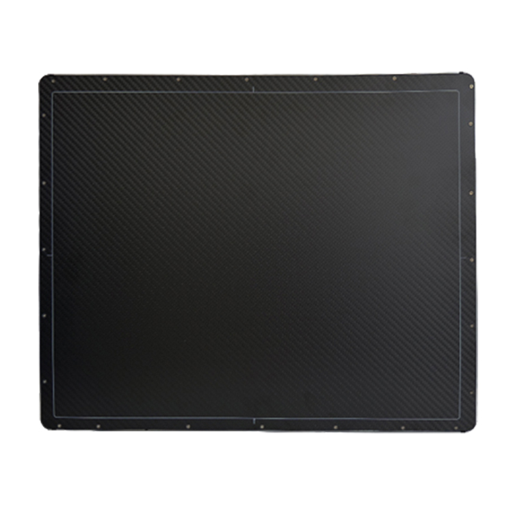
What Is a Mammography Flat Panel Detector?
A flat panel detector (FPD) is the imaging sensor that converts X-rays passing through breast tissue into a digital image. Unlike older computed radiography (CR) systems that use cassettes and phosphor plates, a flat panel detector captures images directly in digital form. This means faster image acquisition, higher contrast resolution, and lower radiation dose — all essential for breast imaging.
There are two main types of flat panel detectors used in mammography:
Direct conversion detectors use amorphous selenium, which converts X-ray photons directly into electrical charge. They offer excellent spatial resolution and are widely preferred for mammography.
Indirect conversion detectors use a scintillator (typically cesium iodide) to convert X-rays into light, which is then detected by a photodiode array. These are common in general radiography and also used in some mammography systems.
For breast imaging specifically, direct conversion detectors are often favored because they deliver the high contrast needed to distinguish subtle differences in soft tissue — a key requirement for spotting microcalcifications and small masses.
Why the Detector Matters for Screening Accuracy
The quality of a mammogram depends heavily on the detector’s performance. Several specifications directly impact clinical outcomes:
Pixel size and spatial resolution. Smaller pixels capture finer detail. A detector with 50–100 μm pixel pitch can resolve microcalcifications as small as 100–200 μm, which are often the earliest sign of breast cancer. Lower-resolution detectors may miss these subtle findings.
Detective quantum efficiency (DQE). DQE measures how efficiently the detector uses the X-ray dose to produce a high-quality image. A higher DQE means better image quality at a lower dose — a critical balance in screening, where women may undergo mammograms annually for decades.
Dynamic range. Breast tissue varies widely in density, from fatty to extremely dense. A detector with a wide dynamic range can capture both very dark and very bright areas in a single exposure without losing detail, reducing the need for repeat imaging.
Artifact performance. Detector defects, such as dead pixels or lines, can mimic or mask pathology. High-quality detectors undergo rigorous quality control and offer long-term stability, minimizing artifacts that could lead to false positives or missed cancers.
Key Features to Look For
When evaluating a mammography flat panel detector for purchase, consider these factors:
Field of view. The detector should cover the full breast, typically 24×30 cm or 24×29 cm. Some systems offer a larger field for imaging larger breasts, reducing the need for multiple exposures.
Pixel pitch. Look for detectors with 50–100 μm pixel size. For screening applications, 70–85 μm is a common sweet spot balancing resolution and dose efficiency.
Dose efficiency. Ask for DQE values at different spatial frequencies. A detector with DQE above 0.5 at low frequencies is generally considered good for mammography.
Compatibility. If you are upgrading an existing mammography system, confirm the detector is compatible with your current generator, gantry, and software. Some detectors are designed as drop-in replacements for specific OEM systems.
Certifications. For clinical use, the detector must meet regulatory standards such as CE marking (Europe), FDA clearance (United States), or equivalent approvals in your region. Verify these before purchase.
Warranty and support. Medical imaging equipment requires ongoing maintenance. Choose a supplier that offers a solid warranty, technical support, and a reliable supply of spare parts.
How to Choose the Right Detector for Your Facility
Start by assessing your current needs:
Screening volume. High-volume screening centers need detectors with fast readout times and high throughput. A detector that can acquire and display an image in under 10 seconds keeps patient flow moving.
Existing equipment. If you are replacing a detector in an existing system, compatibility is the first constraint. Work with the manufacturer or a qualified distributor to confirm fit.
Budget. Direct conversion detectors generally cost more than indirect conversion types but offer superior resolution. Weigh the clinical benefit against your budget.
Future needs. Consider whether you may add tomosynthesis (3D mammography) later. Not all detectors support this mode, so choosing one that does can future-proof your investment.
The flat panel detector is the single most important component in a digital mammography system. It determines image quality, radiation dose, and ultimately the ability to detect breast cancer at its earliest, most treatable stage. When selecting a detector, prioritize spatial resolution, DQE, dynamic range, and regulatory compliance. A well-chosen detector not only improves diagnostic accuracy but also enhances patient comfort through shorter exams and lower dose.
Author: X Ray Machine Accessories
E-mail: newheekcn@163.com
Tel: +86 19015366638
Whatsapp:+86 19015366638
Company: Weifang Newheek Electronic Technology Co., Ltd.
ADD: E Building of Future Star Scientific Innovation Industrial Zone of No.957 Wolong East Street, Yulong Community, Xincheng Sub-District Office, Weifang Hi-tech Zone, Shandong Province, China