In modern veterinary practice, fast and accurate imaging diagnosis is a core necessity to guarantee diagnosis and treatment quality. Whether you run a small community pet clinic or a large 24-hour animal emergency hospital, choosing the right pet X-ray machine can optimize daily diagnosis and treatment workflows, boost diagnostic confidence, and greatly improve the consultation experience for both pets and their owners.
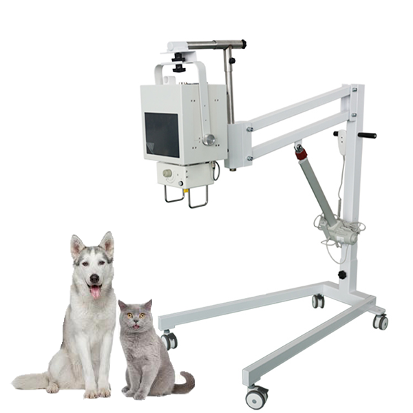
High-definition digital imaging is an essential configuration for modern veterinary clinics
Traditional film-based X-ray systems were once widely used in the veterinary field, but they have inherent drawbacks such as long waiting time for film output, high consumable costs, limited image storage, and blurry details that make it hard to identify tiny lesions. Today’s high-definition digital pet X-ray machine, namely the veterinary DR system, completely breaks these limitations. This pet X-ray machine is equipped with a professional amorphous silicon flat-panel detector with cesium iodide coating, delivering a peak imaging resolution of up to 4.0 lp/mm. It can clearly capture 0.1mm bone cracks, subtle organ nodules, tiny foreign bodies in the digestive tract, and early dental lesions that are easily missed by ordinary devices. It can also present ultra-fine anatomical structures for exotic pets such as hamsters, parrots and rabbits, assisting veterinarians in accurately judging abnormalities that are hard to observe with the naked eye. Unlike old-fashioned devices that require 5-10 minutes of waiting for film development, this digital pet X-ray machine can output high-definition images within 1 second after exposure. For emergency cases such as car accident trauma, accidental foreign body ingestion and severe dyspnea, it eliminates the waiting time for imaging results. You can directly view the images on the 23.8-inch high-definition display, quickly formulate diagnosis and treatment plans, and seize the golden rescue window for critically ill pets.
Low-radiation design protects both pets and clinic staff
Radiation safety is a top concern for many veterinary clinics when selecting X-ray equipment: excessive radiation will bring hidden health risks to young pets, elderly pets, and chronically ill pets that need repeated reexaminations, and will also expose the medical staff who operate the equipment daily to long-term occupational health risks. This high-definition digital pet X-ray machine is equipped with a full-link low-radiation optimization system and a 40kHz high-frequency high-voltage generator, which can precisely control the X-ray energy and dose from the source to avoid unnecessary radiation waste. Combined with the flat-panel detector with ultra-high quantum detection efficiency, it can efficiently capture every weak X-ray signal, and achieve the same or even better imaging effect at a radiation dose far lower than that of traditional X-ray machines. After actual measurement, the single-examination radiation dose of this equipment is only 1/3 of that of traditional DR equipment, far lower than the industry average. Even if pets need multiple reexaminations during the diagnosis and treatment process, it will not cause radiation cumulative damage. At the same time, the equipment adopts a precision shielding design to make the leakage radiation close to zero, which greatly reduces the radiation exposure risk of medical staff during daily operations, allowing the diagnosis and treatment team to focus on their work without worrying about hidden radiation hazards.
Pet-friendly design customized for veterinary scenarios
The pet X-ray machine is not a simple modified version of ordinary medical DR equipment, but is fully customized for the actual diagnosis and treatment scenarios of veterinary clinics. It is pre-installed with an exclusive veterinary image acquisition software, whose parameter setting interface is presented in the form of intuitive animal graphics. Exposure presets for different body types and different examination parts are pre-loaded. Veterinarians only need to select the corresponding pet category and examination site, and the equipment can automatically match the optimal radiation parameters, no repeated manual debugging is required, which greatly reduces the probability of waste films. The radiography bed adopts a four-way floating bed surface with electromagnetic braking, which is convenient for medical staff to quickly fix restless pets. The bed surface is made of medical fiberboard that resists pet scratches, which can keep flat and intact even after long-term use. The equipment supports two exposure modes: bedside touch screen and long-distance remote control. Operators can complete the exposure operation outside the radiation room to further reduce the risk of radiation exposure. In terms of space adaptability, the integrated compact design allows the equipment to be deployed in an area of only about 4 square meters. Even small pet clinics with limited space can complete the layout without large-scale renovation. It supports 220V civilian power supply, no additional circuit transformation is required, and most clinics can complete installation and commissioning shortly after the equipment arrives.
Powerful post-processing functions enable seamless connection with modern clinic systems
This pet X-ray machine fully complies with the DICOM 3.0 international standard, and can be seamlessly connected with the PACS system commonly used in veterinary clinics. All images can be digitally stored, transmitted and backed up, no need to occupy a large amount of space to store physical films, which greatly saves the storage cost and management energy of the clinic. The built-in professional image post-processing toolkit supports multiple operations such as automatic image optimization, edge enhancement and sharpening, zoom roaming, image stitching, window width and window level adjustment, and precise measurement of distance, area and density. Veterinarians can freely adjust image details according to different diagnostic needs: enhance the contrast of bone tissue to observe micro-fractures, optimize soft tissue layering to identify subtle changes in organs, zoom in on the region of interest to check for suspicious lesions. A single X-ray image can release more diagnostic information, helping to find hidden problems that are easily missed in conventional images.
Author: X Ray Machine Accessories
E-mail: newheekcn@163.com
Tel: +86 19015366638
Whatsapp:+86 19015366638
Company: Weifang Newheek Electronic Technology Co., Ltd.
ADD: E Building of Future Star Scientific Innovation Industrial Zone of No.957 Wolong East Street, Yulong Community, Xincheng Sub-District Office, Weifang Hi-tech Zone, Shandong Province, China